Problem Statement:
Oocyte evaluation, beyond nuclear maturity, is challenged by a lack of non-invasive markers of quality – presenting an interesting problem to solve with AI. Model interpretability (understanding how predictions are made) has received increasing attention in medicine. Our aim was to develop and validate an interpretable AI model for oocyte assessments by leveraging image segmentation to predict blastocyst development.
Methods:
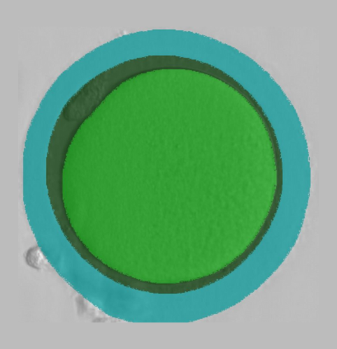
Focusing on 3 specific regions of the oocyte—ooplasm, perivitelline space (PVS), and zona pellucida (ZP)—a Fully Convolutional Branch TransFormer model was developed to segment 2-dimensional images of denuded MII oocytes. Average intersection over union scores were 98.1 ± 0.1%, 97.4 ± 0.6%, and 97.0 ± 0.8% for the ooplasm, PVS, and ZP respectively, indicating accurate segmentation by the model compared to ground truths, as labelled by 3 senior embryologists. A total of 47 features—comprising computations for each region (e.g. circularity), relative geometric features (e.g. ooplasm:ZP area), and cohort-related features—were determined for each sample. Utilizing these features as inputs, a LightGBM classifier model was trained on 40,074 oocyte images with associated laboratory outcomes, to predict blastocyst development. The importance of individual features to model predictions was determined using the Shapley method. The impact of removing features on model performance (in terms of area under the curve; AUC) was also assessed.
Results:
On a test set of 11,757 segmented oocyte images, the blastocyst prediction model displayed an AUC of 0.63 (sensitivity=0.51 and specificity=0.66). The clinical variables—number of MII oocytes and age—were assigned the highest feature importance by the model, but the top ten extracted features corresponded to the ooplasm. Consistent with this observation, removal of ooplasm-related features resulted in a reduction in AUC from 0.63 to 0.57, suggesting its greater relevance to oocyte quality as opposed to extracytoplasmic features (PVS or ZP).
Conclusions:
Accurately segmenting images contributes to developing an interpretable AI model by ensuring exclusive assessment of oocyte features—enhancing trust in the tool. While age is a known predictor of oocyte competence, these results indicate additional measurable features should be considered to better understand oocyte quality.
You Might Also Like …
Join our mailing list for dispatches on the future of fertility